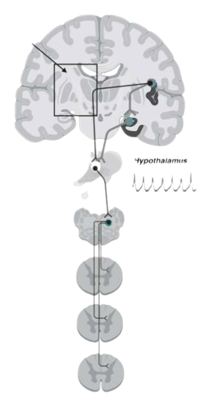
Services
Why we do the work this way.
- Every person is different.
- Every blood pressure is different.
- Every pulse is different.
We take a detailed clinical history and follow how symptoms evolve. We look for patterns of autonomic dysfunction, at rest and under stress. Technological tools for a precise diagnosis, within reach.
Who is this for?
We see children, adolescents, and adults with weakness, lightheadedness, palpitations, fatigue, or poor exercise tolerance. These symptoms — orthostatic intolerance — can reshape life at home, school, or work. The family is part of the evaluation.
01
Tilt-table testing with beat-to-beat monitoring and continuous cerebral blood flow (transcranial Doppler ultrasound)
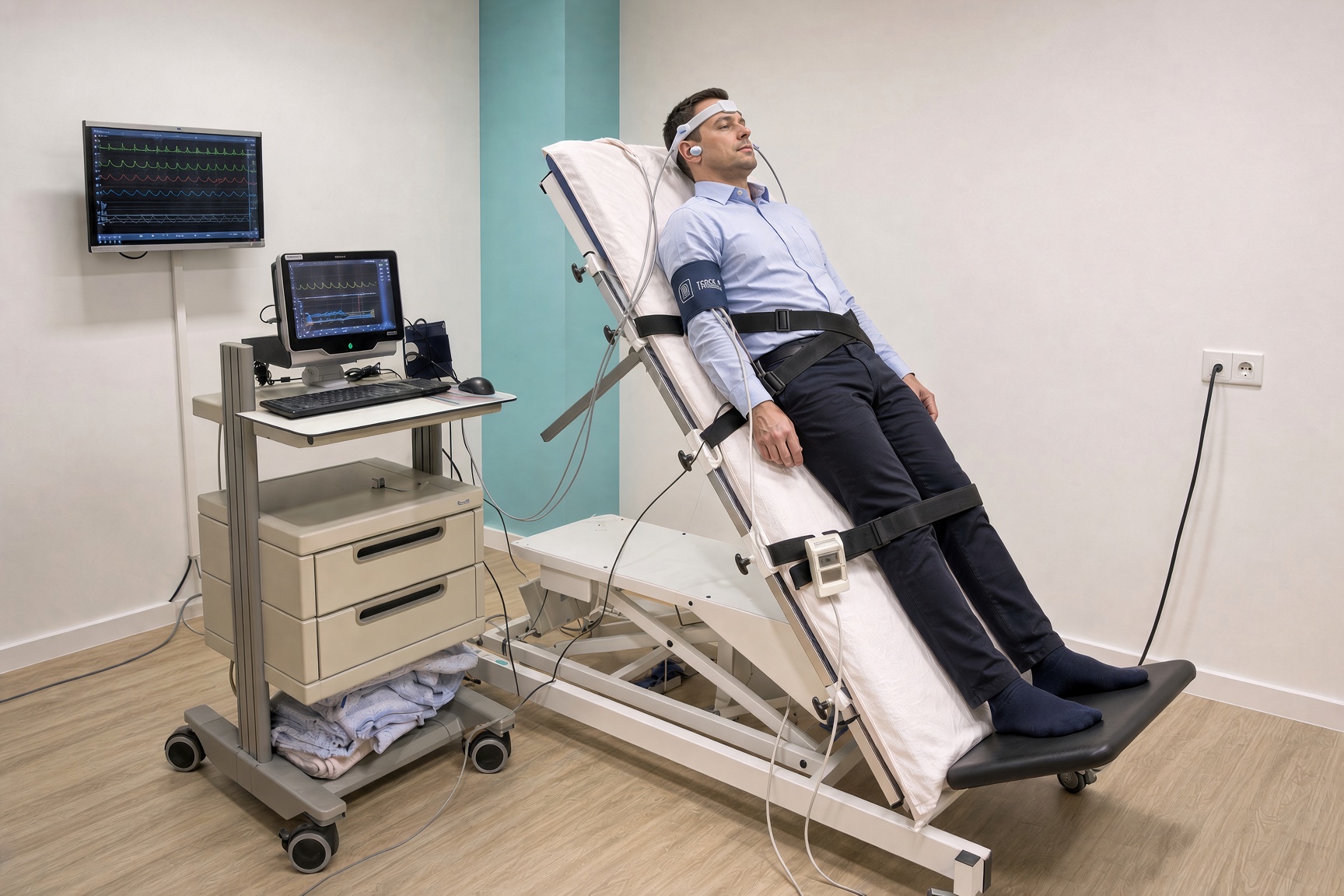
Tilt-table testing shows how your body responds to a change in position, especially from lying down to a controlled 60-degree incline.
You remain on a special table that tilts in a supervised way. We record blood pressure beat-to-beat and, continuously, cerebral blood flow with transcranial Doppler ultrasound.
We perform the study with advanced multi-signal monitoring, in a quiet room, under continuous medical supervision.
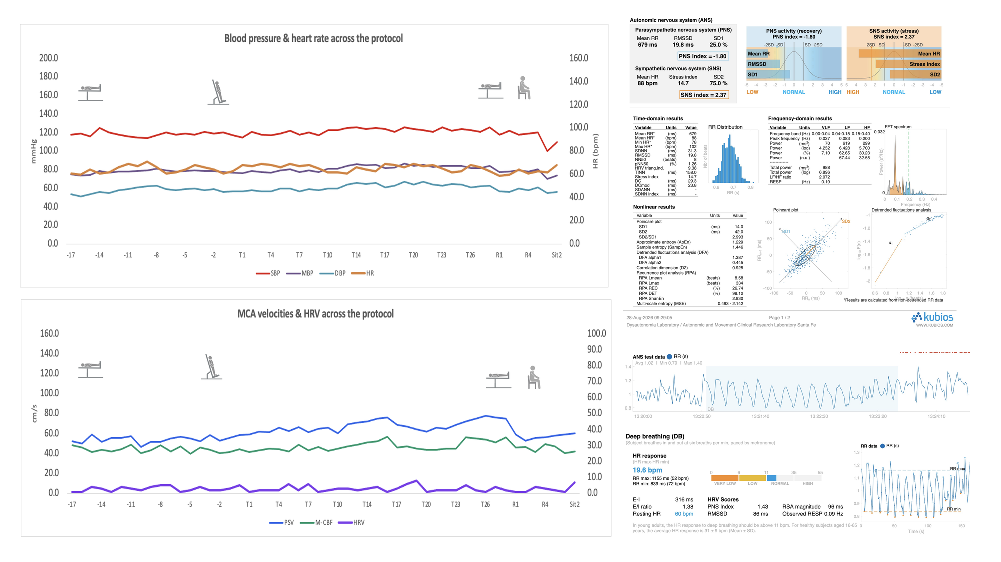
Scroll sideways to see blood pressure, pulse, cerebral flow, and HRV without cropping. Open full size
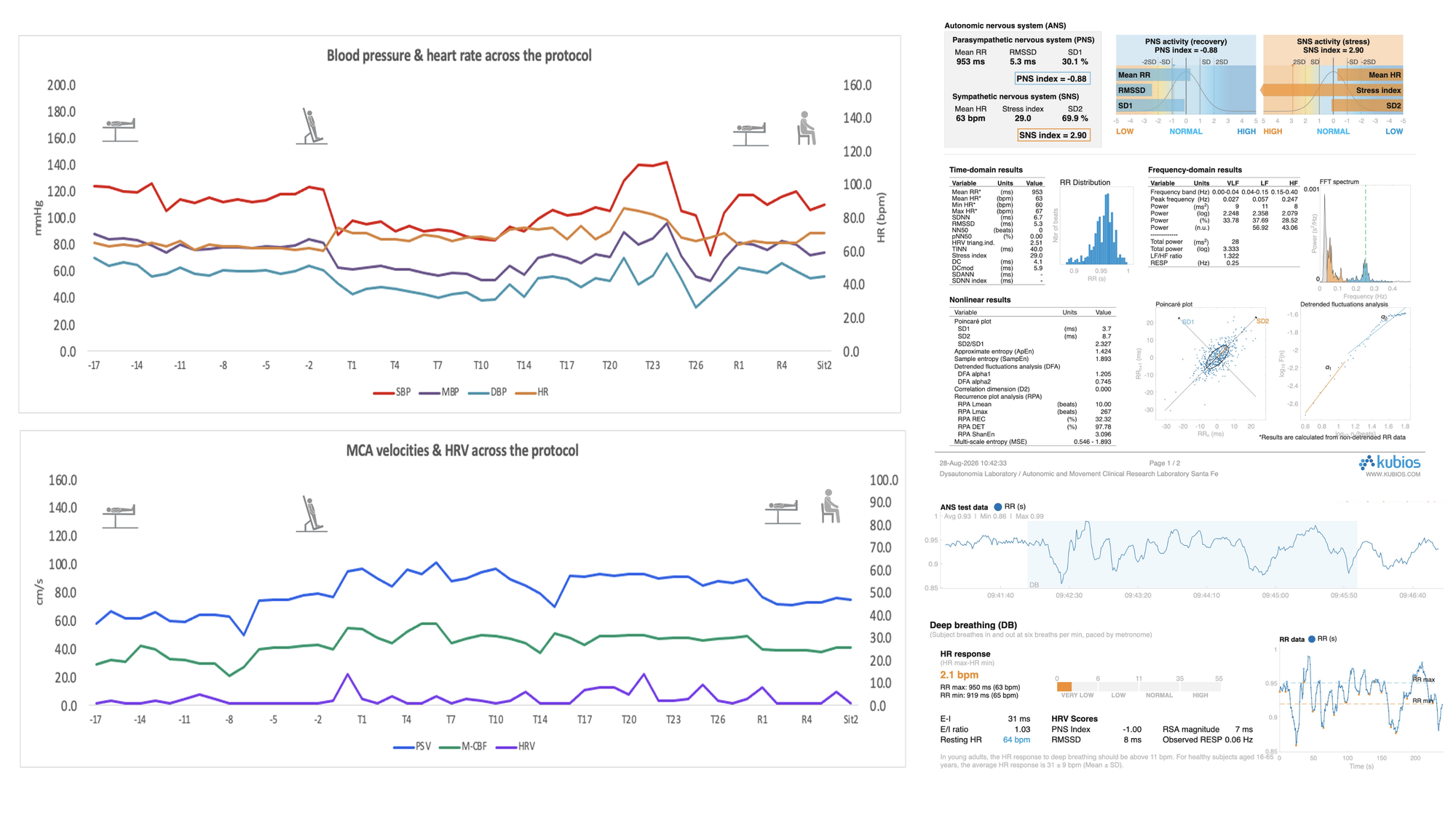
Scroll sideways to see blood pressure, pulse, cerebral flow, and HRV without cropping. Open full size
02
Transcranial ultrasound with a tilt-table protocol for patients with stroke of undetermined etiology (cryptogenic)
A protocol for patients who have already had a stroke whose cause remains unclear — cryptogenic stroke.
We combine transcranial Doppler ultrasound with tilt-table testing to watch how cerebral blood flow changes with posture, and to detect hemodynamic patterns that a resting study can miss.
The report is written for the treating physician and for the etiological work-up of the stroke, with the patient’s consent.
03
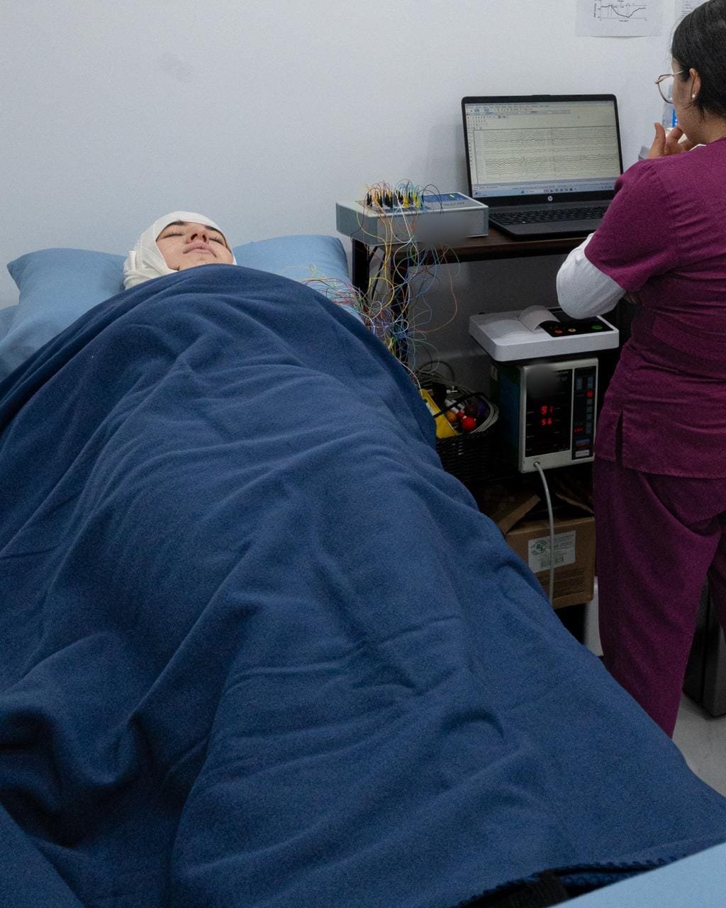
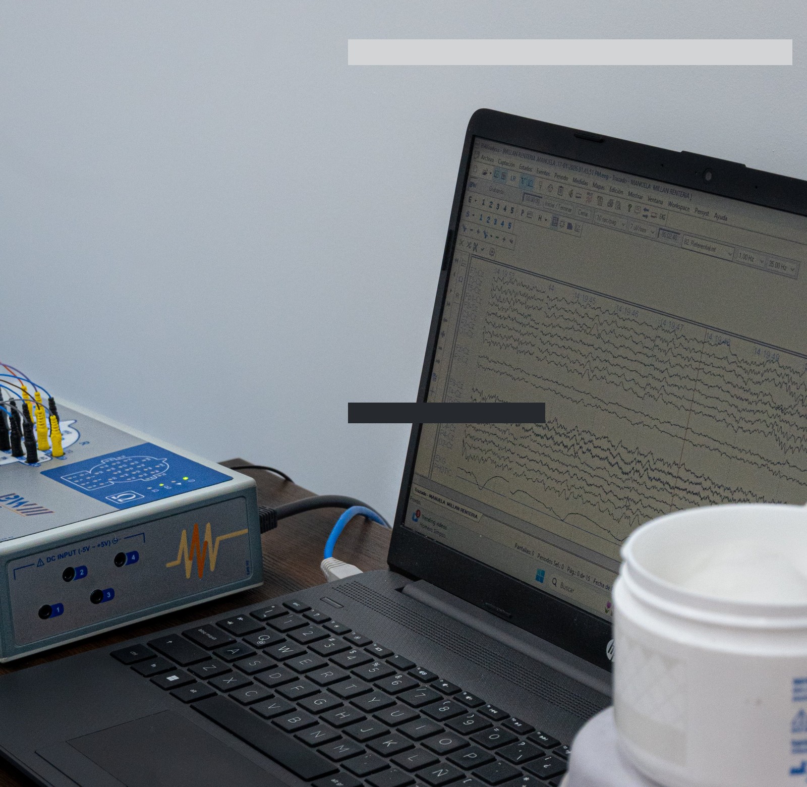
EEG and brain mapping
An electroencephalogram records the brain’s electrical activity through scalp electrodes. It evaluates cerebral function and can detect electrical abnormalities.
Brain mapping analyzes regions in greater detail. It can reveal abnormal patterns in wakefulness and sleep, guide the diagnosis of epilepsy, and help confirm functional non-epileptic events in functional neurological disorders.
When you book, we send the requirements for the study.
04
Cognitive screening
A brief screen for memory, planning, and visuospatial function — often affected in parkinsonian disorders.
Neurogenic hypotension can also change cognition: in severe cases, thinking worsens even while seated. Finding this early lets us adapt treatment.
05
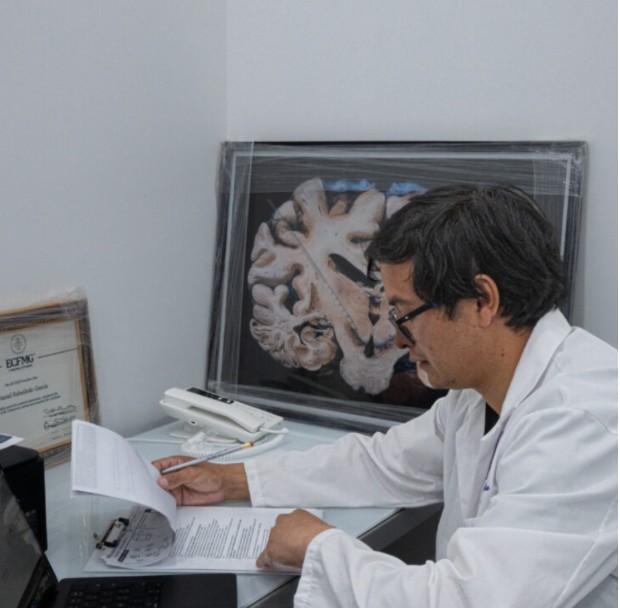
Movement evaluation
A clinical examination of tremor, parkinsonism, ataxia, dystonia, and gait, together with the autonomic symptoms that often travel with them.
It helps distinguish a neurodegenerative disorder from other causes — including functional neurological disorders — and shapes the follow-up plan.
06
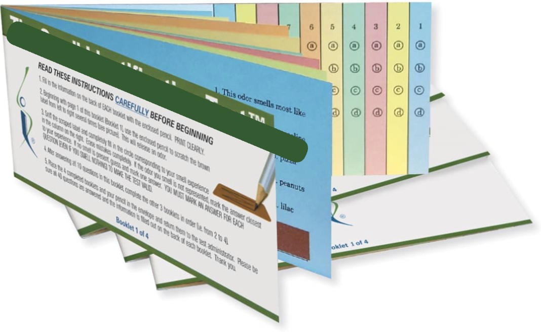
Evaluation of the sense of smell
After COVID-19 in 2020 we learned more about how smell relates to nervous-system disease.
Smell is an ancient sense. The olfactory nerve can be damaged during COVID-19 infection, even in people who had no symptoms.
Conditions such as Parkinson’s disease may begin years or decades before motor signs. Distinguishing them carefully improves treatment and prognosis.
07
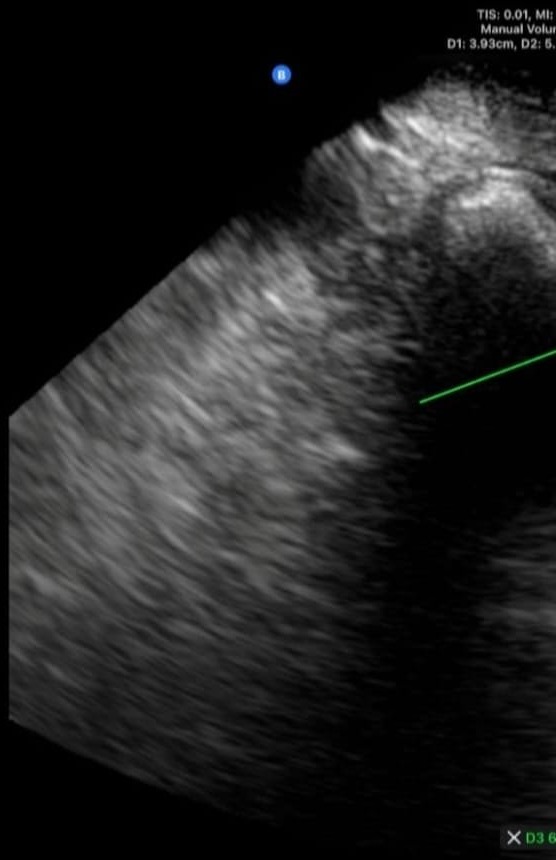
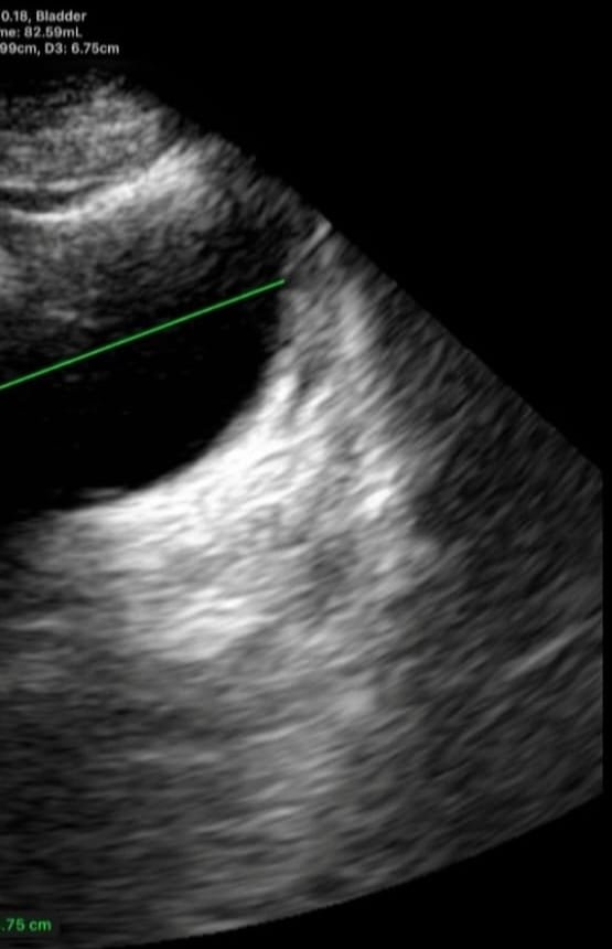
Functional bladder ultrasound
Ultrasound indirectly assesses autonomic (sympathetic and parasympathetic) and somatic nerves that control bladder filling and emptying.
When these nerves fail without structural lesions, the finding is a neurogenic bladder — common in degenerative parkinsonism.
Clinical example: 78-year-old man with degenerative parkinsonism. Pre-void volume 211 ml; post-void 87 ml.
08
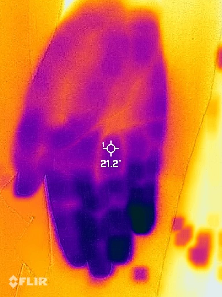
Thermography
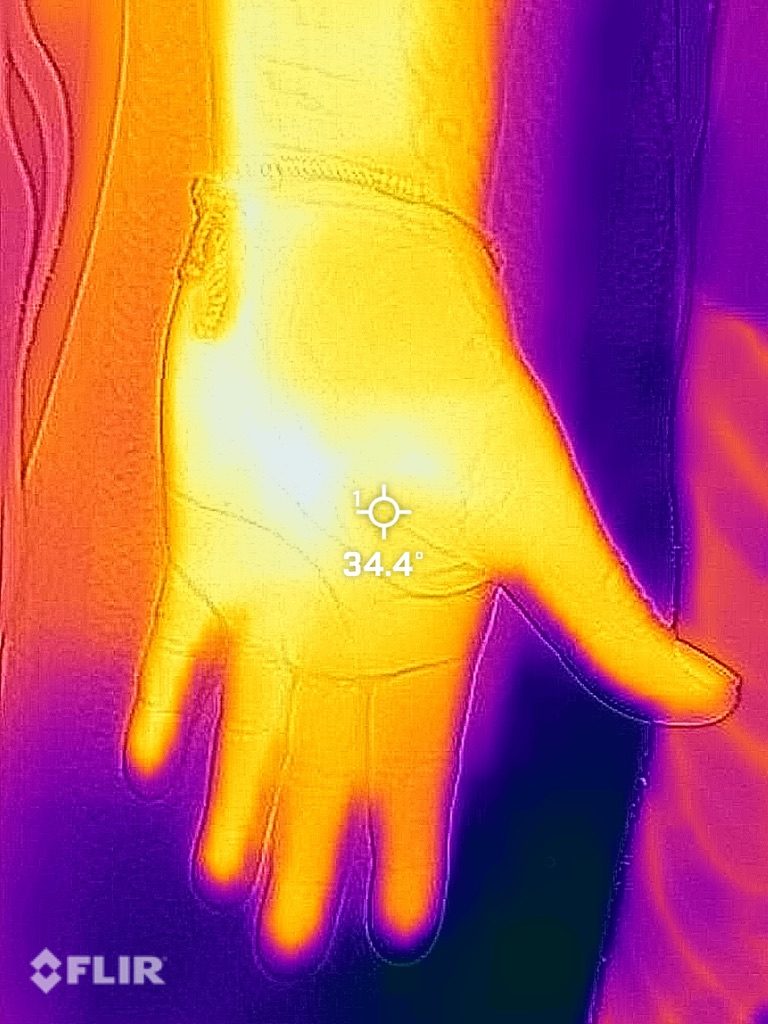
A thermal camera shows how skin temperature changes with posture and in the hands and feet.
In dysautonomia these shifts can appear at rest and during a brief cold-exposure test, adding dynamic information to the autonomic evaluation.
We read the study by comparing a healthy control with a patient with dysautonomia: the thermal pattern of the palms is not the same.
09
Coming soon
Sudomotor evaluation (Sudoscan)
A quantitative measure of the thin nerve fibers that control sweating, using electrochemical skin conductance.
It will complement thermography in the study of small-fiber neuropathy and sudomotor dysautonomia.
Coming soon
10
In-person and virtual visits
Both formats are designed for personal, safe, and convenient care, without geographic barriers.
For first visits, follow-ups, and video calls we ask for enough time to think carefully through your clinical history.